Anatomy Of Upper Thigh And Hip - Hip Anatomy Video Medical Video Library : Several muscles cross the front of the hip and create hip flexion, pulling the thigh and trunk toward both muscles cross the floor of the pelvis, emerge at the outer edges of the pubic bones, and finally insert on the inner upper femur (thighbone).
byAdmin•
0
Anatomy Of Upper Thigh And Hip - Hip Anatomy Video Medical Video Library : Several muscles cross the front of the hip and create hip flexion, pulling the thigh and trunk toward both muscles cross the floor of the pelvis, emerge at the outer edges of the pubic bones, and finally insert on the inner upper femur (thighbone).. The median cubital vein (a common site site for venepuncture) in the antecubital fossa of the arm. Anatomy of the human body. The hip's unique anatomy enables it to be both extremely strong and amazingly flexible, so it can bear weight and allow for a wide range of movement. It is part of the lower limb. Atlas of human anatomy in cross section.
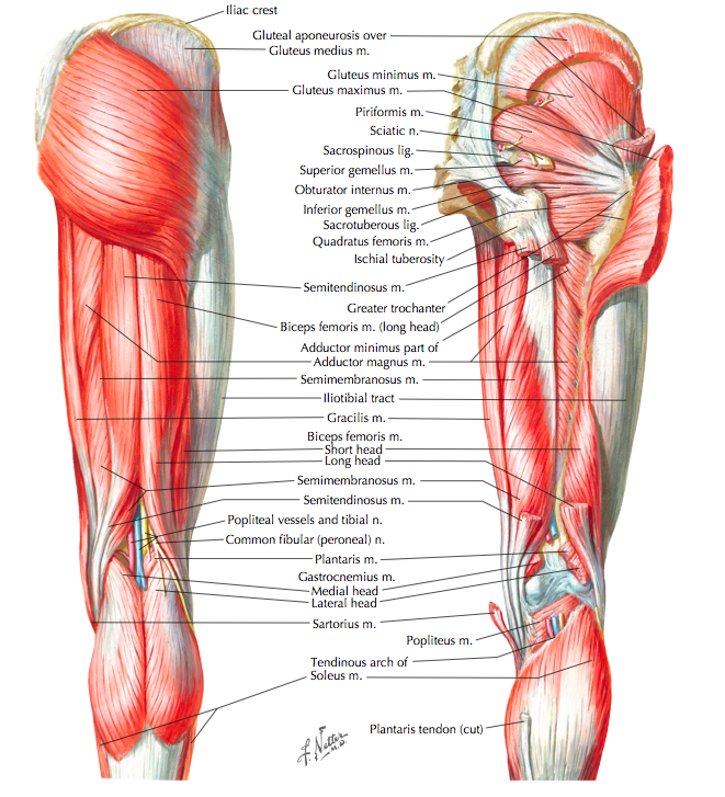
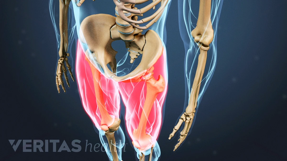
Pelvis, perineum, hip, and upper thigh. Muscles of the hips and thighs. Each pelvic girdle consists of a hip bone (coxal bone, innominate bone), which articulates with the head of a femur. Finally, the hamstring muscles that run down the back of the thigh start on the bottom of the pelvis. The hip joint is a ball and socket joint that is the point of articulation between the head of the femur and the acetabulum of the pelvis.
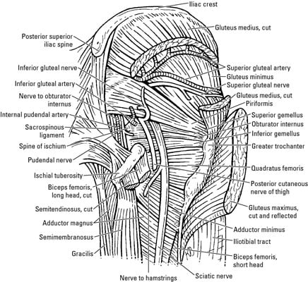
Muscle Synergies Of The Hip And Pelvis Rayner Smale from images.squarespace-cdn.com The iliopsoas muscle, which extends from the lower back to upper femur; The following nerves serve the gluteal and. The muscles also require a lot of blood flow, which provides oxygen and nourishment, especially when you're physically active. While the thigh muscles will be slip into the anterior, medial and posterior groups. Foundational anatomy provides medical students with the necessary background in anatomy for success in clerkships. Chief flexor of knee weak. In vertebrate anatomy, hip (or coxa in medical terminology) refers to either an anatomical region or a joint. The femur or thigh bone is one of the longest bones in the human body.
Hip and knee pain and hip and shoulder pain are.
It's restricted by contact of the thigh with all the abdomen and adduction is restricted by contact. While the thigh muscles will be slip into the anterior, medial and posterior groups. Hip movements include flexion, extension, abduction, adduction, circumduction, and hip rotation. 431).—at the upper and medial part of the thigh, a little below the medial end of the inguinal ligament, is a large. Mri of upper leg (femur). Muscles of the hips and thighs. Medial condyle of tibia nerve supply: Upper part of the ischial tuberosity insertion: Tibial part of the sciatic nerve action: The paired hip bones are connected. The patient lies supine with the hip and knee flexed and the hip externally rotated into the frog leg position. The hip's unique anatomy enables it to be both extremely strong and amazingly flexible, so it can bear weight and allow for a wide range of movement. Knowing the anatomy of your hip can help you understand the source of any hip pain.
This deep muscle begins in the low back and pelvis and connects on the inside edge of the upper femur. The anatomical areas found on the upper limb can serve as key landmarks to help us find important anatomical structures such as finding one of the superficial veins: Several muscles cross the front of the hip and create hip flexion, pulling the thigh and trunk toward both muscles cross the floor of the pelvis, emerge at the outer edges of the pubic bones, and finally insert on the inner upper femur (thighbone). Muscles of the hips and thighs. Upper part of the ischial tuberosity insertion:
Why Do I Feel A Warm Sensation In My Thigh from embed.widencdn.net Atlas of human anatomy in cross section. Chief flexor of knee weak. The following nerves serve the gluteal and. Groin, inguinal region and the anterior and posterior regions of the hip and thigh. A, anterior and posterior views show the hip joint ligaments. Like the forearm, the upper leg, or thigh, has a dense arrangement of many muscles. This mri hip joint axial cross sectional anatomy tool is absolutely free to use. Knowing the anatomy of your hip can help you understand the source of any hip pain.
The thigh is the area between the hip and the knee joint.
The following nerves serve the gluteal and. All of the anatomical parts of the hip work together to enable various movements. The hip's unique anatomy enables it to be both extremely strong and amazingly flexible, so it can bear weight and allow for a wide range of movement. It is part of the lower limb. A, anterior and posterior views show the hip joint ligaments. 340 anatomical structures of the hip region were labeled, accessible on anatomical parts: Finally, the hamstring muscles that run down the back of the thigh start on the bottom of the pelvis. Each pelvic girdle consists of a hip bone (coxal bone, innominate bone), which articulates with the head of a femur. The muscles also require a lot of blood flow, which provides oxygen and nourishment, especially when you're physically active. 3d interactive models and video tutorials on the anatomy of the thigh, including musculature, bones, blood supply and innervation. Medial condyle of tibia nerve supply: The thigh is the area between the hip and the knee joint. Tibial part of the sciatic nerve action:
The cavity of the acetabulum faces obliquely forward, outward, and downward. Groin, inguinal region and the anterior and posterior regions of the hip and thigh. 340 anatomical structures of the hip region were labeled, accessible on anatomical parts: The hip joint is a ball and socket joint that is the point of articulation between the head of the femur and the acetabulum of the pelvis. This webpage presents the anatomical structures found on thigh mri.
Nerves Of The Hip And Thigh Dummies from www.dummies.com Upper part of the ischial tuberosity insertion: Hip and leg pain can cause stress on joints and affect other areas of the body. On the anterior side, the most prominent of the muscles are the sartorius muscle and the four the four muscle of the quadriceps all extend the lower leg, and the rectus femoris additionally can flex the thigh at the hip. It functions to adduct the thigh and to flex. Like the forearm, the upper leg, or thigh, has a dense arrangement of many muscles. Knowing the anatomy of your hip can help you understand the source of any hip pain. Hip and knee pain and hip and shoulder pain are. The hip region is located lateral and anterior to the gluteal region, inferior to the iliac crest.
He also serves the communities of charleston, sc and augusta, ga.
Want to learn more about it? The muscles also require a lot of blood flow, which provides oxygen and nourishment, especially when you're physically active. He also serves the communities of charleston, sc and augusta, ga. Anatomy ▶ lower limb ▶ bones and cartilages ▶ hip joint. Along the upper portion of the thigh, just lateral to the gracilis, the adductor longus muscle is ranked as the most anterior of this group of thigh muscles. Hip surgeon dr guillaume dumont offers hip pain treatments in columbia, sc. It functions to adduct the thigh and to flex. Mri of upper leg (femur). Atlas of human anatomy in cross section. 340 anatomical structures of the hip region were labeled, accessible on anatomical parts: B, muscles of the anterior thigh compartment. The upper part of the thigh bone consists of the femoral head, femoral neck, and greater and lesser trochanters. The different anatomical areas of the gluteal region:
This webpage presents the anatomical structures found on hip mri upper thigh anatomy. The median cubital vein (a common site site for venepuncture) in the antecubital fossa of the arm.